Most patients walk into an eye exam expecting to read letters off a chart and walk out with a new glasses prescription. However, a comprehensive exam covers far more ground than that single test.
Your eye doctor is checking the health of nearly every structure in your eye, and in some cases, picking up early signs of conditions that affect the rest of your body. Keep reading to learn more about what happens during a comprehensive eye exam.
Vision Sharpness and Refractive Errors
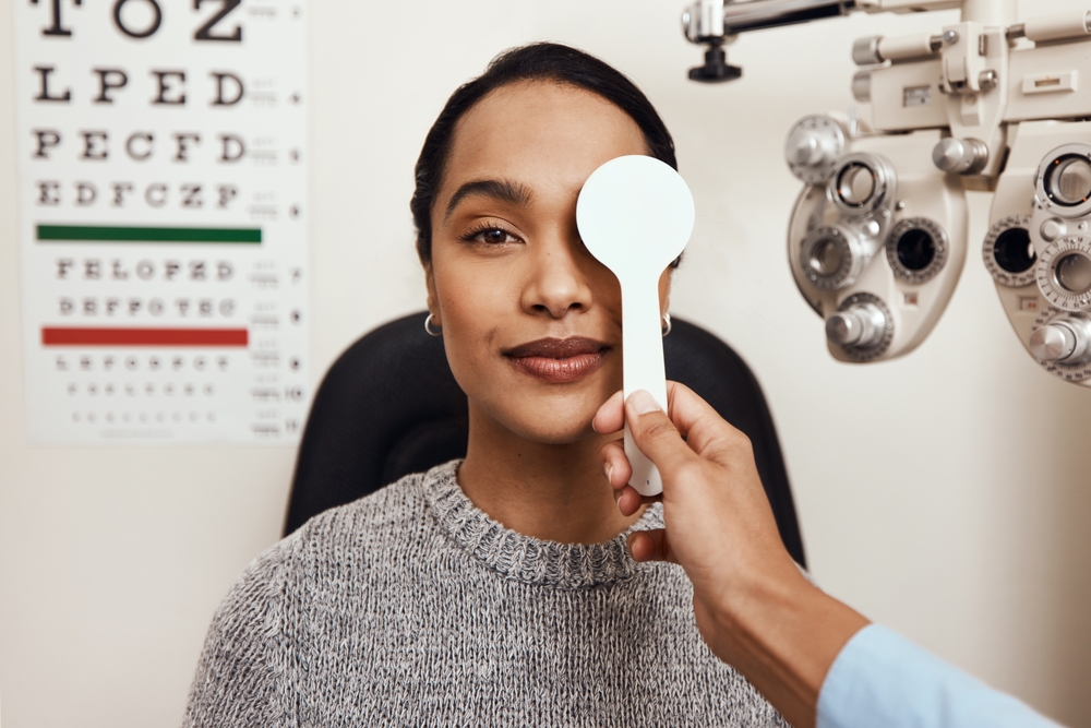
A comprehensive eye exam typically starts with a visual acuity test, the familiar reading of letters at a distance to measure how clearly you see. This establishes a baseline and flags any changes in your vision since your last visit. From there, your eye doctor moves on to refraction testing, switching between lens options to pinpoint the exact prescription that can sharpen your vision.
This step identifies refractive errors such as nearsightedness, farsightedness, and astigmatism, all of which stem from how light bends as it enters the eye. Getting this measurement right is the foundation for accurate glasses or contact lenses.
Eye Muscle Coordination and Movement
Your eye doctor also watches how your eyes move and align with each other. This involves following a light or a moving target while the doctor checks for smooth tracking and proper teamwork between both eyes. Misalignment can cause double vision, eye strain, or a lazy eye that developed in childhood and never fully resolved.
Testing coordination matters even if your vision in each eye tests normally on its own. Two eyes that see clearly individually but do not work well together can still cause headaches, eye strain, or fatigue during reading and screen time.
Children are closely monitored for this, since undetected coordination issues can affect how the brain learns to process input from both eyes. Adults can develop new coordination problems too, particularly after an injury or as certain muscles weaken with age.
Pupil Response
A penlight test checks how your pupils react to light, constricting when light hits them and expanding in dim settings. Sluggish or uneven pupil response can point to neurological issues that extend well beyond the eye itself. Your doctor pairs this with a peripheral vision test to map your full field of sight, not just what is directly in front of you.
Visual Field Testing
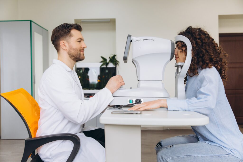
Peripheral vision loss often develops gradually and without obvious symptoms, which is part of why this test matters even for patients who feel their sight is fine. Depending on your age and risk factors, this test might use a simple hand-motion check or a more detailed automated machine that maps blind spots across your entire visual field. Either way, the goal is the same: confirming that your side vision matches what a healthy eye should detect.
Intraocular Pressure
Measuring the pressure inside your eye is one of the more important steps in a comprehensive exam. A quick puff of air or a gentle probe against the eye’s surface gives your eye doctor a pressure reading in millimeters of mercury. Elevated pressure is a primary risk factor for glaucoma, a condition that damages the optic nerve gradually and often without early symptoms.
Because glaucoma can progress for years before a patient notices any vision changes, this pressure check is one of the few ways to catch it while treatment can still preserve sight. Your eye doctor will also look directly at the optic nerve for any changes in its shape or color that might signal damage already underway.
Do you remember when you had your last eye exam?
Slit-Lamp Examination
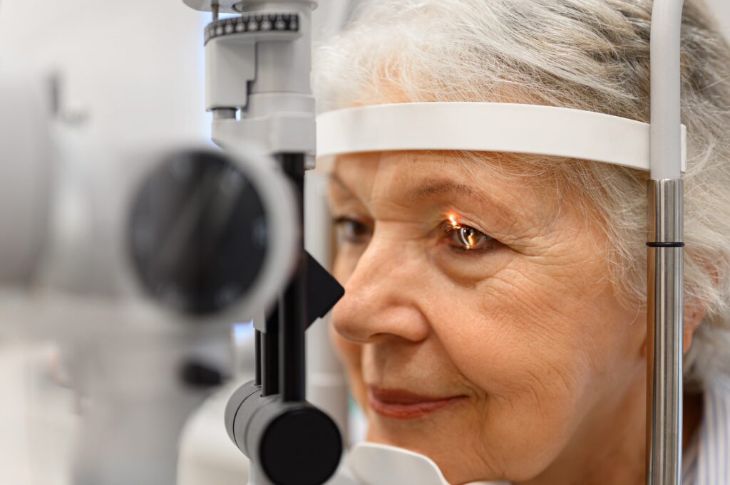
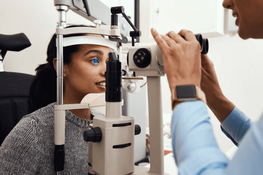
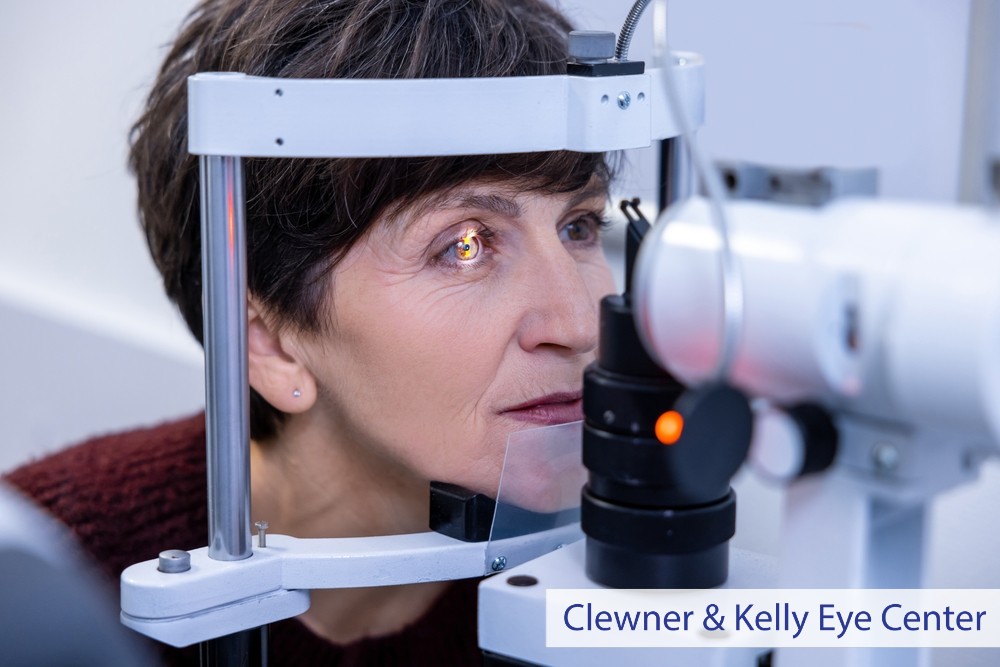
Using a slit-lamp, a microscope with a bright, focused light, your eye doctor examines the cornea, iris, eyelids, and eyelashes in fine detail. This step reveals scratches, infections, inflammation, or structural irregularities that would be invisible during a basic vision check.
Behind the iris sits the eye’s natural lens, and the slit-lamp allows your eye doctor to check its clarity as well. Over time, proteins in the lens can clump together and cloud vision, a process that leads to cataracts. Early cataracts may not affect daily activities yet, but documenting their progression during each exam helps determine the right time for treatment down the road.
The slit-lamp exam also gives your eye doctor a close look at the tear film, which affects comfort and clarity of vision throughout the day.
The Retina and Optic Nerve
The final major step involves dilating your pupils with eye drops so your eye doctor can examine the retina, optic nerve, and blood vessels at the back of your eye. This part of the exam takes longer to set in and leaves your vision blurry and light sensitive for a few hours afterward, but it opens a window that cannot be seen any other way.
Diabetic retinopathy, a complication tied to blood sugar levels, shows up here as changes in the retinal blood vessels, making diabetic eye care a routine part of this evaluation for patients with diabetes.
The dilated exam also screens for macular degeneration, a condition affecting the central part of the retina responsible for sharp, detailed vision. Retinal tears, detachments, and other structural changes are also visible during this portion of the exam.
Because many retinal conditions develop without pain or obvious symptoms, this step catches problems that patients would otherwise not notice until vision loss had already progressed.
Book Your Comprehensive Eye Exam Today
Each piece of a comprehensive exam builds toward a complete picture of your eye health. Pressure readings, retinal images, and lens clarity checks from one visit become the comparison point for the next, allowing your eye doctor to catch subtle changes early.
Some findings, such as changes in blood vessels or the optic nerve, can even reflect conditions that affect your overall health, including diabetes and high blood pressure. Regular comprehensive exams give your eye doctor the history needed to notice small shifts before they become significant problems.
Due for your next comprehensive eye exam? Schedule an appointment at Clewner & Kelly Eye Center in Boca Raton, FL.